Over its nearly 300-year history of development, the microscope has probably become one of the most popular optical devices widely used in all areas of human activity. It is especially difficult to overestimate its role in teaching schoolchildren who know the surrounding microcosm with their own eyes.
A distinctive feature of the proposed microscope is the "non-standard" use of a conventional Web camera. The principle of operation consists in the direct registration of the projection of the studied objects on the surface of the CCD matrix when illuminated by a parallel beam of light. The resulting image is displayed on a PC monitor.
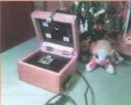
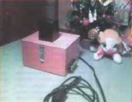
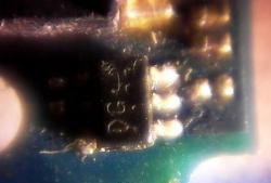
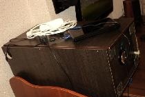
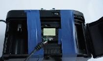
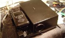
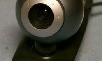
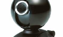
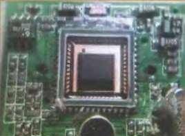
Compared with a conventional microscope, the proposed design lacks an optical system consisting of lenses, and the resolution is determined by the pixel size of the CCD matrix and can reach units of microns. The appearance of the microscope is shown in Fig. 1 and fig. 2. Mustek firm Wcam 300A model was used as a web-camera. It has a color CCD with a resolution of 640x480 pixels. An electronic board with a CCD matrix (Fig. 3) is removed from the case and, after a little refinement, is installed in the center of the lightproof case with an opening lid. The finalization of the board consisted of re-soldering the USB connector in order to provide the possibility of installing additional protective glass on the surface of the CCD matrix and sealing the surface of the board.
A through hole was made in the housing cover, in the center of which there is a block of three LEDs of different glow colors (red, green, blue), which is a light source. The LED block, in turn, is closed by an opaque casing. The remote location of the LEDs from the matrix surface allows the formation of an approximately parallel beam of light at the measurement object.
The CCD is connected to a PC using a USB cable. Software - full-time, included in the delivery of the Web-camera.
The microscope provides an image magnification of 50 ... 100 times, with an optical resolution of about 10 microns with an image refresh rate of 15 Hz.
The design of the microscope is shown in Fig. 4 (not to scale).

For the entrance window of the CCD matrix 7 for its protection against mechanical damage, a quartz protective glass 6 with dimensions 1x15x15 mm was installed. Protection of the electronic board from liquids and mechanical damage is ensured by sealing its surface with silicone sealant 8. The test object 5 is placed on the surface of the protective glass 6. Lighting LEDs 2 are installed in the center of the opening of the lid 4 and are closed externally by a lightproof plastic casing 3. The distance between the test object and the LED block is approximately 50 ... 60 mm.
The power LEDs (Fig. 5) are powered by a battery of 12 out of three 4.5 V. supply voltage. The illumination LEDs EL1 — EL3 are turned on and thereby the lighting color is selected by switches SA2 — SA4 (13) located on the side wall of the housing 11.

Resistors R1, R3 — R5 are current-limiting. Resistor R2 (14) is designed to adjust the brightness of the LEDs EL1 — EL3, it is installed on the rear wall of the housing.The device uses constant resistors C2-23, MLT, variable - SPO, SP4-1. Power switch SA1 - MT1, switches SA2 — SA4 - push-button SPA-101, SPA-102, LED AL307BM can be replaced by KIPD24A-K
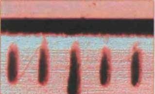
Since the apparent size of the output images depends on the characteristics of the video card used and the size of the monitor, the microscope requires calibration. It consists in registering a test object (transparent school ruler), the dimensions of which are known (Fig. 6). By measuring the distance between the strokes of the ruler on the monitor screen and correlating them with the true size, you can determine the image scale (magnification). In this case, 1 mm of the monitor screen corresponds to 20 μm of the measured object.
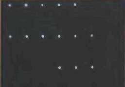
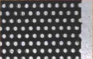
Using a microscope, you can observe various phenomena and measure objects. In fig. 7 shows an image of laser perforation of a banknote of denomination of 500 rubles. The average diameter of the holes is 100 μm, the scatter of the holes in shape is visible. In fig. 8 is an image of a Hitachi color picture mask mask. The diameter of the holes is about 200 microns.
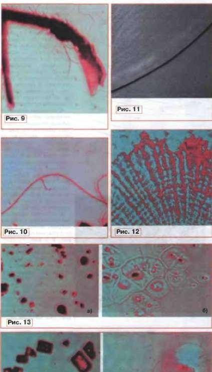
As an example of biological objects, a spider, its paw and mustache are selected; they are shown in fig. 9 and fig. 10, respectively (the diameter of the mustache is about 40 microns), the hair of the author (diameter - 80 microns) - in Fig. 11, fish scales - in fig. 12. It is interesting to observe the processes of dissolution of substances in water. As an example, the processes of dissolution of salt and sugar are given. In fig. 13a and fig. Fig. 14a shows particles of dry salt and sugar crystals, respectively, and Fig. 13.6 and fig. 14.6 - the process of their dissolution in water. Zones of increased concentration of substances and the effects of focusing light at the centers of dissolution are clearly visible.
Source: Radio 1`2008